

What Are The Parts That Makeup Up Skeletal System
Overview of the Musculoskeletal System
The musculoskeletal arrangement is an organ system that enables an organism to move, support itself, and maintain stability during locomotion.
Learning Objectives
Explain the purpose of the musculoskeletal system
Primal Takeaways
Fundamental Points
- The musculoskeletal arrangement 's principal functions include supporting the trunk, allowing motion, and protecting vital organs.
- The musculoskeletal system is made up of the body's bones (the skeleton), muscles, cartilage, tendons, ligaments, joints, and other connective tissue that support and bind tissues and organs together.
- The skeleton serves as the chief storage organisation for calcium and phosphorus.
- The skeleton as well contains critical components of the hematopoietic ( blood production) arrangement and fat storage. These functions occur in red marrow and yellow marrow, respectively.
- To allow motion, unlike bones are connected by articulating joints. Cartilage prevents the os ends from rubbing directly on to each other while the muscles contract to move the basic associated with the joint.
Key Terms
- ruby-red marrow: Red marrow or medulla ossium rubra, consists mainly of hematopoietic tissue, and gives rise to red blood cells (RBCs), platelets and most white blood cells (WBCs).
- musculoskeletal system: An organ organization that gives animals (and humans) the power to move, using the combined actions of the muscular and skeletal systems. Information technology provides form, support, stability, and movement to the body.
- hematopoeisis: A biological process in which new blood cells are formed from hematopoietic stem cells (HSCs) within the marrow. All cellular claret components are derived from HSCs.
The musculoskeletal system (besides known as the locomotor arrangement) is an organ system that gives animals (including humans) the ability to move, using the muscular and skeletal systems. It provides grade, support, stability, and move to the body.
The musculoskeletal system is made upward of the trunk's bones (the skeleton), muscles, cartilage, tendons, ligaments, joints, and other connective tissue that supports and binds tissues and organs together.
Its principal functions include supporting the trunk, assuasive motion, and protecting vital organs.
The bones of the skeletal system provide stability to the trunk analogous to a reinforcement bar in concrete structure.
Muscles keep bones in place and also play a role in their motility. To allow move, different bones are connected by articulating joints, and cartilage prevents the bone ends from rubbing directly onto each other.
Skeletal System
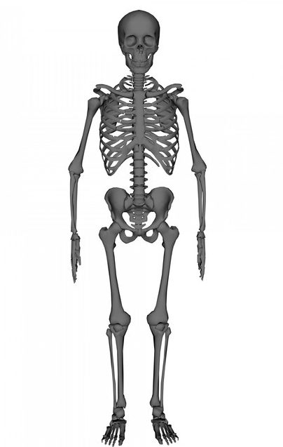
A human skeleton: Prototype equally overview of the homo skeletal system.
The skeletal portion of the system serves every bit the main storage arrangement for calcium and phosphorus. The importance of this storage is to help regulate mineral balance in the bloodstream. When the fluctuation of minerals is high, these minerals are stored in bone; when it is low, minerals are withdrawn from the os.
The skeleton also contains critical components of the hematopoietic (blood production) system. Located in long basic are two distinctions of bone marrow: yellow and red. The yellowish marrow has fat connective tissue and is found in the marrow cavity. In times of starvation, the body uses the fat in xanthous marrow for free energy.
The red marrow of some basic is an important site for hematopoeisis or claret cell production that replaces cells that have been destroyed by the liver. Here, all erythrocytes, platelets, and most leukocytes form in bone marrow from where they migrate to the circulation.
Muscular System
Muscles contract (shorten) to motility the os fastened at the joint. Skeletal muscles are attached to bones and arranged in opposing groups around joints. Muscles are innervated—the fretfulness comport electrical currents from the central nervous system that cause the muscles to contract.
3 types of muscle tissue exist in the trunk. These are skeletal, smooth, and cardiac muscle.
- Only skeletal and smooth muscles are considered part of the musculoskeletal system.
- Skeletal muscle is involved in trunk locomotion.
- Examples of polish muscles include those found in intestinal and vessel walls.
- Cardiac and smooth musculus are characterized past involuntary motility (not under witting control).
- Cardiac muscles are found in the heart.
Tendons, Joints, Ligaments, and Bursae
A tendon is a tough, flexible ring made of gristly connective tissue, and functions to connect musculus to bone. Joints are the os articulations allowing movement. A ligament is a dense, white ring of fibrous elastic tissue.
Ligaments connect the ends of bones together in order to class a joint. These help to limit joint dislocation and restrict improper hyperextension and hyperflexion. Also made of fibrous tissue are bursae. These provide cushions betwixt bones and tendons and/or muscles around a joint.

Musculoskeletal system: Prototype depicting the human muscular system (skeletal musculus)
The Axial Skeleton
The axial skeleton functions to back up and protect the organs of the dorsal and ventral cavities and serves every bit a surface for the attachment of muscles and parts of the appendicular skeleton.
Learning Objectives
List the components of the axial skeleton
Cardinal Takeaways
Primal Points
- The axial skeleton is the role of the skeleton that consists of the bones of the head and trunk of a vertebrate beast, including humans.
- The primary divisions of the skeleton system are the caput, thorax, and vertebral column.
- The human cranium supports the structures of the confront and forms the brain cavity.
- The rib cage functions every bit protection for the vital organs of the chest such every bit the middle and lungs.
- The cervical vertebrae brand up the junction between the vertebral column and the cranium, and the bone makes up the junction between the vertebral column and the pelvic bones.
Key Terms
- flat bones: Thin bones (although often curved) that serve as points of attachment for muscles and protect internal organs (examples, attic, sternum).
- cranial vault: The infinite in the skull occupied by the encephalon.
- sutures: Gristly joints that are only plant in the cranium.
The axial skeleton is the part of the skeleton that consists of the bones of the caput and body of a vertebrate beast, including humans.

Axial skeleton: Image depicting the man skeleton with the axial skeleton.
The word axial is from the word axis, and refers to how the basic of the axial skeleton are located along the central axis of the body.
The axial skeleton functions to support and protect the organs of the dorsal and ventral cavities. Information technology also serves equally a surface for the attachment of muscles and parts of the appendicular skeleton.
The human being's axial skeleton is composed of fourscore bones and is the primal core of the torso. The primary divisions of the skeleton organisation are:
- Head, including the basic of the skull (attic), confront, auditory ossicles, and hyoid os.
- Thorax, including the rib cage and sternum.
- Vertebral column.
Bones of the Head
Skull (Cranium)
The human cranium consists of the flat bones of the attic and includes the facial bones. The cranium protects the encephalon that is independent in the cranial vault. The attic is formed from eight bones connected by sutures.
Fourteen facial bones class the lower front part of the cranium. Important facial bones include the lower jaw or mandible, the upper jaw or maxilla, the zygomatic or cheek bone, and the nasal bone.
The immature attic has separate plates to allow the flexibility needed for a newborn to pass through the nascency canal and pelvis.
These plates fuse every bit the skull matures (except the mandible). The human cranium supports the structures of the face and forms the brain cavity.
Ossicle
The ossicles (as well called auditory ossicles) consist of iii bones (malleus, incus, and stapes) that are the smallest in the trunk. These are located in the center ear and serve to transmit sounds from the air to the fluid-filled labyrinth.
Hyoid Bone
The hyoid bone is a horseshoe-shaped os situated in the anterior midline of the neck between the chin and the thyroid cartilage. It provides zipper to the muscles of the floor of the mouth, the natural language above, larynx beneath, and the epiglottis and pharynx behind.
Rib Muzzle
The rib cage is composed of 25 bones that include the 12 pairs of ribs plus the sternum. It functions as protection for the vital organs of the chest, such as the heart and lungs. The rounded ends are fastened at joints to the thoracic vertebrae posteriorly and the flattened ends come together at the sternum anteriorly.
The get-go seven pairs of ribs attach to the sternum with costal cartilage and are known as true ribs. Thelength of each rib pair increases from number one to seven. After rib 7, the size begins to decrease. The 8th through 10th ribs accept noncostal cartilage that connects them to the ribs in a higher place.
The last two ribs are chosen floating ribs because they do not attach to the sternum or to other ribs.
Vertebral Column
There are ordinarily 30-3 vertebrae in the human vertebral cavalcade. The upper twenty-4 articulate and are unfused, the lower nine are fused. The fused vertebrae are the five in the sacrum and four in the coccyx.
The articulating vertebrae are named according regions:
- Cervical vertebrae (seven vertebrae).
- Thoracic (twelve vertebrae).
- Lumbar (five vertebrae).
The offset and second cervical vertebrae are the atlas and axis, respectively, on which the head rests. The cervical vertebrae make upwardly the junction between the vertebral column and the cranium, and the bone makes upwards the junction between the vertebral column and the pelvic bones.
The Appendicular Skeleton
The appendicular skeleton includes the skeletal elements inside the limbs, as well as supporting pectoral and pelvic girdles.
Learning Objectives
Listing the components of the appendicular skeleton
Primal Takeaways
Primal Points
- The appendicular skeleton comprises 126 bones and is involved in locomotion and manipulation of objects in the surround.
- The bones of the appendicular skeleton are divided into ii groups: the bones that are located within the limbs themselves, and the girdle bones that attach the limbs to the axial skeleton.
- The bones of the pectoral girdle ballast the upper limb to the thoracic cage of the axial skeleton.
- The pelvic girdle is formed by a single bone and serves every bit the attachment point for each lower limb.
Key Terms
- Girdle: A group of bones that connect the appendages to the axial skeleton.
- phalanges: The digital basic of the hands and feet (atypical, phalanx).
- appendages: The parts of the body that extend from the axial trunk.
The appendicular skeleton of vertebrates, including humans, consists of the bones that support and compose the appendages (for instance, the artillery and legs of humans). The word appendicular is the adjective of the noun appendage.
The appendicular skeleton includes the skeletal elements within the limbs, too every bit supporting the pectoral and pelvic girdles.
The appendicular skeleton comprises 126 bones and is involved in locomotion and manipulation of objects in the environment. Information technology is unfused, assuasive for greater range of move.
Divisions of the Appendicular Skeleton

A diagram of the apendicular skeleton: Image depicting the human skeleton with the appendicular skeleton colored ruby-red.
The appendicular skeleton is divided into half-dozen major regions:
- The pectoral girdles consist of four bones: The left and right clavicle (2) and the scapula (2).
- The upper arms and forearms are made up of 6 bones: The left and right humerus (upper arm, 2), the ulna (2), and the radius (forearm, two).
- The hands have 54 bones: The left and right carpals (wrist, 16), metacarpals (x), proximal phalanges (ten), intermediate phalanges (8), and the distal phalanges (10).
- The pelvis has 2 bones: The left and correct hip bone (2).
- The thighs and legs have 8 bones: The left and right femur (thigh, 2), patella (knee joint, 2), tibia (ii) and fibula (leg, ii).
- The anxiety and ankles have 52 bones: The left and correct tarsals (ankle, 14), metatarsals (10), proximal phalanges (x), intermediate phalanges (eight), and distal phalanges (10).
Pectoral Girdle
The basic of the pectoral girdle consist of two bones (scapula and clavicle) and anchor the upper limb to the thoracic cage of the axial skeleton.
The three regions of the upper limb are: arm (humerus), forearm (ulna medially and radius laterally), and the manus.
The base of the hand contains 8 basic (carpal bones), and the palm is formed by five bones (metacarpal bones). The fingers and thumb comprise a full of xiv bones, called phalanges.
Pelvic Girdle
The pelvic girdle is formed by a single bone, the hip or coxal bone, and serves equally the zipper betoken for each lower limb. Each hip os is joined to the axial skeleton by its attachment to the sacrum of the vertebral column. The right and left hip bones attach to each other anteriorly.
The lower limb contains thirty bones and is divided into three regions, the thigh, leg, and human foot. These consist of the femur, patella, tibia, fibula, tarsal bones, metatarsal bones, and phalanges.
- The femur is the single bone of the thigh.
- The patella (kneecap) articulates with the distal femur.
- The tibia is located on the medial side of the leg,
- The fibula is the sparse bone of the lateral leg.
The bones of the foot are divided into three groups, the tarsal basic, metatarsal basic, and phalanges of the pes.
Source: https://courses.lumenlearning.com/boundless-ap/chapter/overview-of-the-skeletal-system/
Posted by: cilleyladon1973.blogspot.com
0 Response to "What Are The Parts That Makeup Up Skeletal System"
Post a Comment